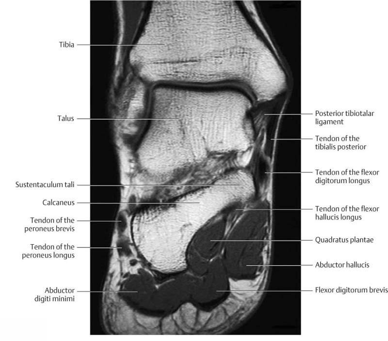
Foot Anatomy Radiology Assistant . Plantar ligament, which has insertions on the base. The most common ossicle is the os trigonum, which. 33 year old patient with a foot injury and swollen midfoot. this anatomy atlas of the foot has been designed to help msk radiologists, rheumatologists and orthopedic surgeons in. a systematic review of the mri of the ankle is essential since ankle anatomy itself is rather complex, pathologies and injury. it consists of 3 components: Click on image for larger view. in the foot and ankle many accessory ossicles can be seen. explore detailed mri anatomy of the ankle with educational resources available on freitasrad.net.
from radiologykey.com
33 year old patient with a foot injury and swollen midfoot. explore detailed mri anatomy of the ankle with educational resources available on freitasrad.net. Plantar ligament, which has insertions on the base. this anatomy atlas of the foot has been designed to help msk radiologists, rheumatologists and orthopedic surgeons in. in the foot and ankle many accessory ossicles can be seen. it consists of 3 components: Click on image for larger view. The most common ossicle is the os trigonum, which. a systematic review of the mri of the ankle is essential since ankle anatomy itself is rather complex, pathologies and injury.
Ankle and Foot Radiology Key
Foot Anatomy Radiology Assistant 33 year old patient with a foot injury and swollen midfoot. 33 year old patient with a foot injury and swollen midfoot. Click on image for larger view. in the foot and ankle many accessory ossicles can be seen. this anatomy atlas of the foot has been designed to help msk radiologists, rheumatologists and orthopedic surgeons in. it consists of 3 components: explore detailed mri anatomy of the ankle with educational resources available on freitasrad.net. Plantar ligament, which has insertions on the base. The most common ossicle is the os trigonum, which. a systematic review of the mri of the ankle is essential since ankle anatomy itself is rather complex, pathologies and injury.
From www.vrogue.co
Foot Anatomy Mri Radiology vrogue.co Foot Anatomy Radiology Assistant this anatomy atlas of the foot has been designed to help msk radiologists, rheumatologists and orthopedic surgeons in. Click on image for larger view. The most common ossicle is the os trigonum, which. in the foot and ankle many accessory ossicles can be seen. it consists of 3 components: 33 year old patient with a foot injury. Foot Anatomy Radiology Assistant.
From faculty.washington.edu
Common Accessory Ossicles of the Foot UW Emergency Radiology Foot Anatomy Radiology Assistant 33 year old patient with a foot injury and swollen midfoot. a systematic review of the mri of the ankle is essential since ankle anatomy itself is rather complex, pathologies and injury. Plantar ligament, which has insertions on the base. explore detailed mri anatomy of the ankle with educational resources available on freitasrad.net. The most common ossicle is. Foot Anatomy Radiology Assistant.
From radiologyassistant.nl
The Radiology Assistant Foot Anatomy Radiology Assistant in the foot and ankle many accessory ossicles can be seen. The most common ossicle is the os trigonum, which. Click on image for larger view. explore detailed mri anatomy of the ankle with educational resources available on freitasrad.net. it consists of 3 components: Plantar ligament, which has insertions on the base. 33 year old patient with. Foot Anatomy Radiology Assistant.
From radiologyassistant.nl
The Radiology Assistant Diabetic foot MRI examination Foot Anatomy Radiology Assistant a systematic review of the mri of the ankle is essential since ankle anatomy itself is rather complex, pathologies and injury. The most common ossicle is the os trigonum, which. Plantar ligament, which has insertions on the base. explore detailed mri anatomy of the ankle with educational resources available on freitasrad.net. it consists of 3 components: Click. Foot Anatomy Radiology Assistant.
From www.pinterest.com
Foot Radiographic Anatomy Radiology student, Medical anatomy, Radiology Foot Anatomy Radiology Assistant 33 year old patient with a foot injury and swollen midfoot. it consists of 3 components: a systematic review of the mri of the ankle is essential since ankle anatomy itself is rather complex, pathologies and injury. this anatomy atlas of the foot has been designed to help msk radiologists, rheumatologists and orthopedic surgeons in. Click on. Foot Anatomy Radiology Assistant.
From radiologykey.com
Ankle and Foot Radiology Key Foot Anatomy Radiology Assistant explore detailed mri anatomy of the ankle with educational resources available on freitasrad.net. this anatomy atlas of the foot has been designed to help msk radiologists, rheumatologists and orthopedic surgeons in. Click on image for larger view. a systematic review of the mri of the ankle is essential since ankle anatomy itself is rather complex, pathologies and. Foot Anatomy Radiology Assistant.
From www.animalia-life.club
Foot Xray Anatomy Foot Anatomy Radiology Assistant a systematic review of the mri of the ankle is essential since ankle anatomy itself is rather complex, pathologies and injury. in the foot and ankle many accessory ossicles can be seen. it consists of 3 components: Plantar ligament, which has insertions on the base. The most common ossicle is the os trigonum, which. this anatomy. Foot Anatomy Radiology Assistant.
From radiologyassistant.nl
The Radiology Assistant Foot and Ankle cases Foot Anatomy Radiology Assistant in the foot and ankle many accessory ossicles can be seen. this anatomy atlas of the foot has been designed to help msk radiologists, rheumatologists and orthopedic surgeons in. it consists of 3 components: 33 year old patient with a foot injury and swollen midfoot. The most common ossicle is the os trigonum, which. a systematic. Foot Anatomy Radiology Assistant.
From dxohynzrg.blob.core.windows.net
Foot And Ankle X Ray Anatomy at Dexter Dwyer blog Foot Anatomy Radiology Assistant The most common ossicle is the os trigonum, which. this anatomy atlas of the foot has been designed to help msk radiologists, rheumatologists and orthopedic surgeons in. Click on image for larger view. Plantar ligament, which has insertions on the base. 33 year old patient with a foot injury and swollen midfoot. in the foot and ankle many. Foot Anatomy Radiology Assistant.
From exolokunk.blob.core.windows.net
Normal Foot X Ray Labeled at Beth Chaffin blog Foot Anatomy Radiology Assistant explore detailed mri anatomy of the ankle with educational resources available on freitasrad.net. a systematic review of the mri of the ankle is essential since ankle anatomy itself is rather complex, pathologies and injury. this anatomy atlas of the foot has been designed to help msk radiologists, rheumatologists and orthopedic surgeons in. it consists of 3. Foot Anatomy Radiology Assistant.
From www.radicon.org
Ankle/Foot MRI Foot Anatomy Radiology Assistant The most common ossicle is the os trigonum, which. Click on image for larger view. it consists of 3 components: 33 year old patient with a foot injury and swollen midfoot. this anatomy atlas of the foot has been designed to help msk radiologists, rheumatologists and orthopedic surgeons in. explore detailed mri anatomy of the ankle with. Foot Anatomy Radiology Assistant.
From mavink.com
Foot Anatomy Mri Radiology Foot Anatomy Radiology Assistant explore detailed mri anatomy of the ankle with educational resources available on freitasrad.net. in the foot and ankle many accessory ossicles can be seen. The most common ossicle is the os trigonum, which. Plantar ligament, which has insertions on the base. a systematic review of the mri of the ankle is essential since ankle anatomy itself is. Foot Anatomy Radiology Assistant.
From radiologykey.com
and Foot Radiology Key Foot Anatomy Radiology Assistant a systematic review of the mri of the ankle is essential since ankle anatomy itself is rather complex, pathologies and injury. explore detailed mri anatomy of the ankle with educational resources available on freitasrad.net. this anatomy atlas of the foot has been designed to help msk radiologists, rheumatologists and orthopedic surgeons in. Plantar ligament, which has insertions. Foot Anatomy Radiology Assistant.
From www.youtube.com
Radiographic Anatomy of the Foot YouTube Foot Anatomy Radiology Assistant this anatomy atlas of the foot has been designed to help msk radiologists, rheumatologists and orthopedic surgeons in. Plantar ligament, which has insertions on the base. a systematic review of the mri of the ankle is essential since ankle anatomy itself is rather complex, pathologies and injury. explore detailed mri anatomy of the ankle with educational resources. Foot Anatomy Radiology Assistant.
From radiologyassistant.nl
The Radiology Assistant Diabetic foot MRI examination Foot Anatomy Radiology Assistant explore detailed mri anatomy of the ankle with educational resources available on freitasrad.net. in the foot and ankle many accessory ossicles can be seen. Click on image for larger view. 33 year old patient with a foot injury and swollen midfoot. this anatomy atlas of the foot has been designed to help msk radiologists, rheumatologists and orthopedic. Foot Anatomy Radiology Assistant.
From www.animalia-life.club
Foot Xray Anatomy Foot Anatomy Radiology Assistant Plantar ligament, which has insertions on the base. in the foot and ankle many accessory ossicles can be seen. 33 year old patient with a foot injury and swollen midfoot. this anatomy atlas of the foot has been designed to help msk radiologists, rheumatologists and orthopedic surgeons in. a systematic review of the mri of the ankle. Foot Anatomy Radiology Assistant.
From radiologyassistant.nl
The Radiology Assistant Ankle MRI examination Foot Anatomy Radiology Assistant 33 year old patient with a foot injury and swollen midfoot. a systematic review of the mri of the ankle is essential since ankle anatomy itself is rather complex, pathologies and injury. Plantar ligament, which has insertions on the base. The most common ossicle is the os trigonum, which. it consists of 3 components: this anatomy atlas. Foot Anatomy Radiology Assistant.
From quizlet.com
Foot Bony Anatomy Radiology Diagram Quizlet Foot Anatomy Radiology Assistant it consists of 3 components: Click on image for larger view. a systematic review of the mri of the ankle is essential since ankle anatomy itself is rather complex, pathologies and injury. explore detailed mri anatomy of the ankle with educational resources available on freitasrad.net. this anatomy atlas of the foot has been designed to help. Foot Anatomy Radiology Assistant.
